Amyloid ÃÆà ½Ãâò2-Microglobulin
Yoshihiro Motomiya and Yuichiro Higashimoto
DOI10.21767/2472-5056.100062
Yoshihiro Motomiya1* and Yuichiro Higashimoto2
1Suiyukai Clinic, Kashihara, Nara, Japan
2Department of Chemistry, Kurume University School of Medicine, Kurume, Fukuoka, Japan
- *Corresponding Author:
- Yoshihiro Motomiya
Suiyukai Clinic, 676-1 Kuzumoto-cho, Kashihara, Nara, 6340007, Japan
E-mail: motomiya@silver.ocn.ne.jp
Received date: May 24, 2018; Accepted date: June 11, 2018; Published date: June 15, 2018
Citation: Motomiya Y, Higashimoto Y (2018) Amyloid β2-Microglobulin. J Clin Exp Nephrol Vol 3:11. doi: 10.21767/2472-5056.100062
Copyright: © 2018 Motomiya Y, et al. This is an open-access article distributed under the terms of the Creative Commons Attribution License, which permits unrestricted use, distribution, and reproduction in any medium, provided the original author and source are credited.
Abstract
Dialysis-related amyloidosis (DRA), which is an inevitable complication of long-term haemodialysis (HD), manifests major clinic-pathological characteristics, including carpal tunnel syndrome, which is the most common clinical sign; systemic involvement of many articular tissues; and the presence of β2-microglobulin (β2M) as a precursor protein in this amyloidosis.
Keywords
Dialysis-related amyloidosis; β2-microglobulin; Haemodialysis
Introduction
Amyloid is a fibrillar protein and is primarily a conformational variant of an originally globular precursor protein. β2M is an essential protein for normal conformation of major histocompatibility complex class I molecules on cell membranes and is degraded mainly in renal proximal tubules. β2M passes freely across vascular walls. Therefore, serum levels of β2M are extremely high in patients undergoing HD. Although HD can clear as much as 70% of circulating β2M, β2M in the interstitial space can accumulate and progressively lead to development of DRA [1]. The amyloid processes may occur in the interstitial space. We have studied the amyloidogenicity of β2M and found a previously unknown amyloidogenic process, which we describe in this short review.
Specific information about the different β2M structures follows here:
The three-dimensional structure (conformation) of β2M: β2M consists of 99 amino acids. The three-dimensional structure of native β2M is formed by the folding of seven peptide segments (strands) into a globular conformation, with both the N-terminal and the C-terminal segments folded inward from the molecular surface. McParland et al. demonstrated that unfolding in β2M started first in the N-terminal segment and then continued in the C-terminal segment [2].
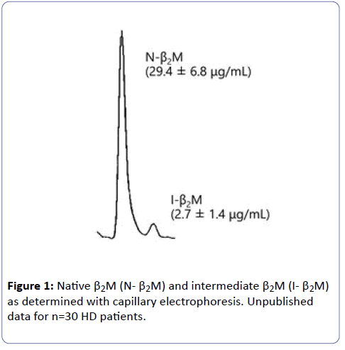
The intermediate structure of β2M: Several basic studies described a conversion process in which amyloid protein is formed. In this process, the folded conformation, i.e., native β2M is unfolded into a partially unfolded conformer, i.e., the intermediate species. The amyloid protein is insoluble, but the intermediate species is soluble and can be identified directly, with native β2M, via capillary electrophoresis. By using this technique [3], we confirmed that β2M in serum consisted of two forms, the folded native β2M (N-β2M) and the partially unfolded intermediate β2M (I-β2M) in both healthy persons and patients undergoing HD (Figure 1) [4,5].
C-terminal unfolding and β2M 92-99: Stoppini et al. first implicated the C-terminal region of β2M in amyloidogenicity by using a monoclonal antibody specific for the C-terminal eight amino acids [6].We then reported on β2M with an unfolded C-terminal region, i.e., β2M 92-99, in amyloid tissues from patients undergoing HD [7]. However, we did not detect β2M 92-99 in serum from patients undergoing HD because the C-terminal region in I-β2M may be not completely unfolded as is the case for β2M 92-99 [8].
ΔN6β2M: In 1987, Linke et al. first reported a variant of β2M lacking the six N-terminal amino acids, i.e., ΔN6β2M, in patients undergoing HD [9]. Esposito et al. and others then confirmed that ΔN6β2M was a highly amyloidogenic variant [10,11]. We recently reported that the C-terminal region was completely unfolded in ΔN6β2M as well as in β2M 92-99 [8].
Glycosaminoglycans (GAGs) and heparin: GAGs including heparin are an essential component of interstitial tissue. In addition, heparin is most often used as an anti-coagulant in the clinical setting of HD. Several studies showed that GAGs facilitate the amyloidogenicity of β2M [12,13], and we also found that heparin promoted β2M unfolding [14].
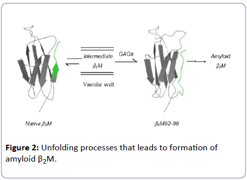
The working hypothesis: N-β2M coexists with I-β2M at a ratio of 10:1 in body fluids, and the HD procedure causes a shift from N-β2M toward I-β2M. GAGs in the interstitial space promote unfolding of the C-terminal region of I-β2M to generate β2M 92-99. β2M 92-99 cannot be refolded or returned to the vascular space. An accumulation of β2M 92-99 leads to the generation of amyloid β2M in the interstitial space (Figure 2).
ΔN6 β2M aptamer: We recently showed that an aptamer for ΔN6 β2M has a domain for the C-terminal region and could block amyloid fibril formation in-vitro [15].
Conclusion
Amyloidosis, including DRA and Alzheimer disease, is becoming a major clinical entity. Amyloidosis is a disease of misfolded precursor proteins. Thus far, we have demonstrated that misfolding of β2M is initiated by an unfolding of the C-terminal segment of β2M, and GAGs in interstitial tissue are intimately associated with the completion of the C-terminal unfolding of β2M. We believe that an intermediate β2M with an unfolded C-terminal may be a key intermediate molecule for amyloid β2M, as (Figure 2) illustrates. The data related to the ΔN6 β2M aptamer support our belief.
References
- Strihou YVC, Floege J, Jadoul M (1994) Amyloidosis and its relationship to different dialysis. Nephrol Dial Transplant 9: 156-161.
- McParland VJ, Kalverda AP, Homans SW, Radford SE (2002) Structural properties of an amyloid precursor of β2-microglobulin. Nat Struct Biol 9: 326-331.
- Heegaard NH, Sen JW, Nissen MH (2000) Congophilicity (Congo red affinity) of different β2-microglobulin conformations characterized by dye affinity capillary electrophoresis. J Chromatogr A 894: 319-327.
- Uji Y, Motomiya Y, Ando Y (2009) A circulating β2-microglobulin intermediate in hemodialysis patients. Nephron Clin Pract111: c173-c181.
- Motomiya Y, Uji Y, Ando Y (2012) Capillary electrophoretic profile of β2-microglobulin intermediate associated with hemodialysis. Ther Apher Dial 16: 350-354.
- Stoppini M, Bellotti V, Mangione P, Ferri G (1997) Use of anti- β2-microglobulin mAb to study formation of amyloid fibrils. Eur J Biochem 249: 21-26.
- Motomiya Y, Ando Y, Haraoka K, Sun X, Morita H, et al. (2005) Studies on unfolded β2-microglobulin at C-terminal in dialysis-related amyloidosis. Kidney Int 67: 314-320.
- Motomiya Y, Higashimoto Y, Uji Y, Suenaga G, Ando Y (2015) C-terminal unfolding of an amyloidogenic β2-microglobulin fragment: DN6 β2M-microglobulin. Amyloid 22: 54-60.
- Linke RP, Hampl H, Lobeck H, Ritz E, Bommer J, et al. (1989) Lysine-specific cleavage of β2-microglobulin in amyloid deposits associated with hemodialysis. Kidney Int 36: 675-681.
- Esposito G, Michelutti R, Verdone G, Viglino P, Hernández H, et al. (2009) Removal of the N-terminal hexapeptide from human β2-microglobulin facilitates protein aggregation and fibril formation. Protein Sci 9: 831-845.
- Myers SL, Jones S, Jahn TR, Morten JI, Tennent GA, et al. (2006) A systematic study of the effect of physiological factors on β2-microglobulin amyloid formation at neutral pH. Biochem 45: 2311-2321.
- Yamamoto S, Yamaguchi I, Hasegawa K, Tsutsumi S, Goto Y, et al. (2004) Glycosaminoglycans enhance the trifluoroethanol-induced extension of β2-microglobulin-related amyloid fibrils at a neutral pH. J Am Soc Nephrol 15: 126-133.
- Borysik AJ, Morten IJ, Radford SE, Hewitt EW (2007) Specific glycosaminoglycans promote unseeded amyloid formation from β2-microglobulin under physiological conditions. Kidney Int 72: 174-181.
- Fukasawa K, Higashimoto Y, Motomiya Y, Uji Y, Ando Y (2016) Influence of heparin molecular size on the induction of C-terminal unfolding in β2-microglobulin. Mol Biol Res Commun 5: 225-232.
- Fukasawa K, Higashimoto Y, Ando Y, Motomiya Y (2017) Selection of DNA aptamer that blocks the fibrillogenesis of a proteolytic amyloidogenic fragment β2M. Ther Apher Dial 22: 61-66.
Open Access Journals
- Aquaculture & Veterinary Science
- Chemistry & Chemical Sciences
- Clinical Sciences
- Engineering
- General Science
- Genetics & Molecular Biology
- Health Care & Nursing
- Immunology & Microbiology
- Materials Science
- Mathematics & Physics
- Medical Sciences
- Neurology & Psychiatry
- Oncology & Cancer Science
- Pharmaceutical Sciences


